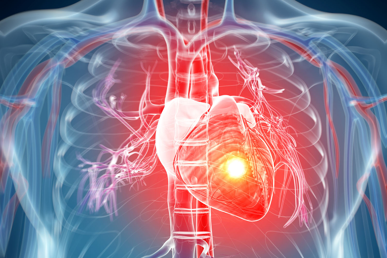
Overview
The anatomy of the heart is a marvel of biological engineering, intricately designed to sustain life by pumping blood throughout the body. Comprising a network of chambers, valves, vessels, and specialized tissues, the heart orchestrates the continuous circulation of oxygen-rich blood to nourish tissues and organs while simultaneously removing metabolic waste products. Let’s delve into the essential components that constitute the anatomy of the heart:
Chambers:
The heart consists of four chambers: two atria (singular: atrium) and two ventricles. The right atrium receives deoxygenated blood returning from the body via the superior and inferior vena cavae, while the left atrium receives oxygenated blood from the lungs via the pulmonary veins.
Blood flows from the atria into the ventricles through atrioventricular (AV) valves, namely the tricuspid valve on the right side and the mitral (or bicuspid) valve on the left side.
Valves:
Valves within the heart ensure the unidirectional flow of blood. The tricuspid valve regulates blood flow between the right atrium and right ventricle, while the mitral valve governs flow between the left atrium and left ventricle.
Semilunar valves, including the pulmonary valve and aortic valve, guard the exits of the ventricles, preventing the backflow of blood into the ventricles after each contraction.
Vessels:
Coronary arteries supply the heart muscle with oxygenated blood. The left coronary artery branches into the left anterior descending artery and the circumflex artery, while the right coronary artery nourishes the right atrium and ventricle.
Coronary veins collect deoxygenated blood from the heart muscle and drain it into the coronary sinus, which empties into the right atrium.
Conduction System:
The heart’s conduction system coordinates its rhythmic contractions. The sinoatrial (SA) node, located in the right atrium, serves as the heart’s natural pacemaker, initiating electrical impulses that spread through the atria and stimulate atrial contraction.
The impulses then travel to the atrioventricular (AV) node, located near the junction of the atria and ventricles, where they are briefly delayed before passing through the bundle of His and Purkinje fibers to stimulate ventricular contraction.
Pericardium:
The heart is enveloped by a double-layered sac called the pericardium. The fibrous pericardium provides structural support and anchors the heart within the chest cavity, while the serous pericardium secretes fluid to lubricate the heart’s movements and reduce friction.
Understanding the intricacies of the anatomy of the heart is fundamental to comprehending its function and pathology. From the rhythmic pulsations of its chambers to the precise coordination of its conduction system, the heart embodies the exquisite synergy of form and function, sustaining life with unwavering dedication and precision.