Overview
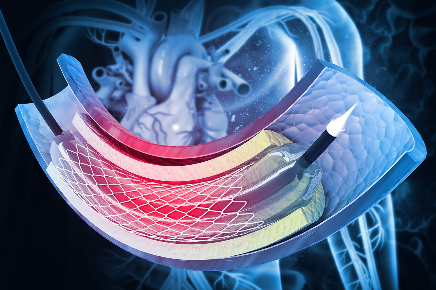
An angiogram, also known as an angiography, is a medical imaging technique used to visualize the inside of blood vessels and organs of the body, particularly the arteries, veins, and the heart chambers. This procedure plays a crucial role in diagnosing and managing cardiovascular diseases, which remain a leading cause of morbidity and mortality worldwide.

What is an Angiogram?
An angiogram involves injecting a contrast dye into the blood vessels and capturing X-ray images to detect any blockages, narrowing, or other abnormalities. The resulting images help doctors assess the condition of the blood vessels and plan appropriate treatments, such as angioplasty or surgery.
The Procedure
The angiogram procedure typically follows these steps:
Preparation: The patient may need to fast for several hours before the procedure. Pre-procedure tests may include blood tests and a physical examination.
Injection of Contrast Dye: A catheter is inserted into a large blood vessel, usually in the groin or arm. Through this catheter, a contrast dye is injected.
Imaging: As the dye flows through the blood vessels, X-ray images are taken. These images reveal the structure and function of the vessels.
Post-Procedure: The catheter is removed, and pressure is applied to the insertion site to prevent bleeding. Patients are monitored for a few hours for any adverse reactions.
Applications of Angiograms
Angiograms are vital in diagnosing various cardiovascular conditions, including:
Coronary Artery Disease (CAD): Angiograms can identify blockages in the coronary arteries, which supply blood to the heart. This information is critical for planning interventions like stenting or bypass surgery.
Aneurysms: By highlighting the blood vessels, angiograms can reveal aneurysms—abnormal bulges in the vessel wall that can rupture if left untreated.
Peripheral Artery Disease (PAD): This procedure helps in diagnosing blockages in the arteries of the legs, which can lead to pain and mobility issues.
Stroke: Angiograms can detect blockages or malformations in the cerebral arteries, aiding in the prompt treatment of strokes.
Advancements in Angiography
Technological advancements have significantly improved the safety and efficacy of angiograms. Modern techniques such as Digital Subtraction Angiography (DSA) enhance image clarity by subtracting pre-contrast images from post-contrast images, reducing the amount of dye needed and minimizing radiation exposure.
Additionally, the development of non-invasive alternatives like CT angiography and MR angiography has provided options for patients who may not be suitable candidates for traditional angiograms due to risks associated with invasive procedures.
Risks and Considerations
While angiograms are generally safe, they do carry some risks, including:
Allergic Reactions: Some patients may be allergic to the contrast dye.
Bleeding or Bruising: At the catheter insertion site.
Kidney Damage: In patients with pre-existing kidney conditions, the contrast dye may exacerbate kidney problems.
Radiation Exposure: Although minimal, repeated exposure can pose long-term risks.
Angiograms are indispensable tools in modern medicine, providing detailed and accurate images of blood vessels that guide the diagnosis and treatment of numerous cardiovascular conditions. Ongoing research and technological innovations continue to enhance the safety, accuracy, and accessibility of angiography, ultimately improving patient outcomes worldwide.
For anyone facing cardiovascular issues, understanding the role and process of an angiogram can help in making informed decisions about their healthcare. As always, discussing with a healthcare professional is crucial to understand the benefits and risks specific to individual health needs.


