Overview
In the realm of medicine, abbreviations and acronyms play a significant role in streamlining communication among healthcare professionals. One such acronym is ECO, which stands for “Echocardiography.” This diagnostic tool is crucial in the field of cardiology and offers detailed insights into the structure and function of the heart.
What is Echocardiography?
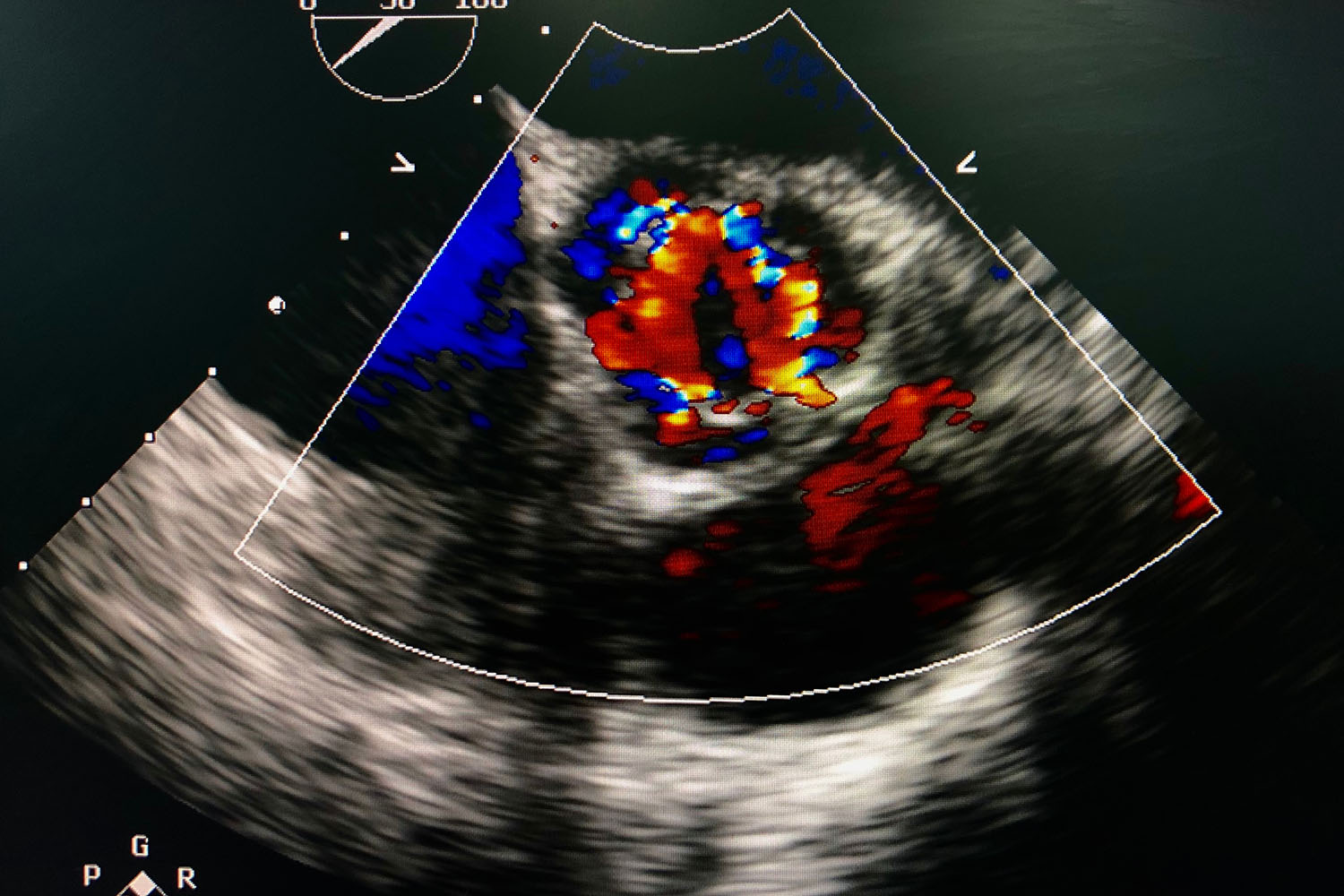
Echocardiography (ECO full form in medical terminology) is a non-invasive imaging technique that uses ultrasound waves to produce images of the heart. These images, called echocardiograms, help doctors evaluate the heart’s size, shape, and motion, as well as its chambers, valves, and surrounding structures.
How Does Echocardiography Work?
Echocardiography employs high-frequency sound waves that are transmitted through a device called a transducer. The transducer is placed on the patient’s chest and sends sound waves into the heart. These waves bounce off the heart’s structures and return to the transducer, which then converts them into electrical signals. These signals are processed by a computer to create real-time images of the heart.
Types of Echocardiography
There are several types of echocardiography, each serving a specific purpose:
1. Transthoracic Echocardiography (TTE):
- The most common type, where the transducer is placed on the chest wall.
- It provides images of the heart’s chambers, valves, and blood flow.
2. Transesophageal Echocardiography (TEE):
- Involves inserting a specialized transducer into the esophagus.
- Offers clearer images of the heart, especially the back structures, as it avoids the interference of the chest wall and lungs.
3. Stress Echocardiography:
- Conducted during or after physical exercise or medication that increases heart rate.
- Helps evaluate how the heart performs under stress and can detect conditions like coronary artery disease.
4. Doppler Echocardiography:
- Uses Doppler signals to assess blood flow and pressure within the heart.
- Essential for identifying issues such as valve problems and congenital heart defects.
Importance of Echocardiography in Medical Diagnosis
Echocardiography (ECO full form in medical diagnostics) is indispensable for diagnosing and managing various heart conditions. It provides comprehensive information about:
- Heart Structure: Detects abnormalities in the heart’s anatomy, such as septal defects or hypertrophy.
- Valve Function: Evaluates the performance of heart valves and identifies issues like stenosis or regurgitation.
- Blood Flow: Assesses the direction and speed of blood flow through the heart and major vessels, helping in the diagnosis of conditions like heart failure or pulmonary hypertension.
- Heart Function: Measures the heart’s pumping efficiency, including ejection fraction, which is critical for diagnosing and monitoring heart failure.
International Research and Advancements
Research and technological advancements continue to enhance the capabilities and applications of echocardiography. International studies have contributed significantly to improving the accuracy and utility of this diagnostic tool.
For instance, advancements in 3D echocardiography provide more detailed and accurate images, aiding in better diagnosis and treatment planning. Research in strain imaging, a technique that assesses the deformation of heart muscle fibers, offers deeper insights into myocardial function, especially in conditions like cardiomyopathies.
Understanding the ECO full form in medical contexts is crucial for appreciating its role in cardiac care. Echocardiography is a vital diagnostic tool that provides detailed information about the heart’s structure and function, helping in the diagnosis, treatment, and management of various heart conditions. As research and technology continue to evolve, echocardiography will undoubtedly become even more integral to cardiovascular medicine, ensuring better patient outcomes worldwide.


