Overview
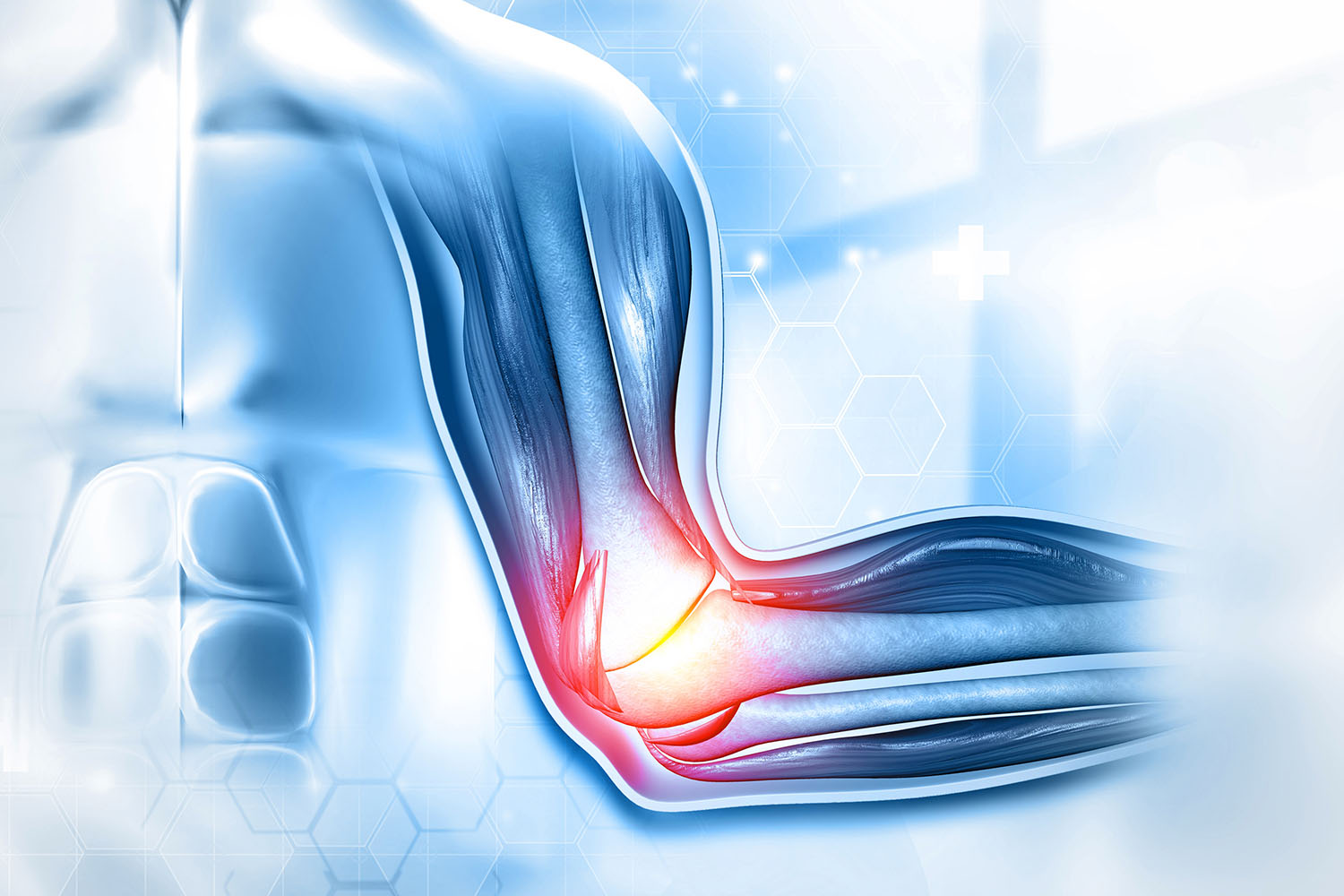
The elbow is a hinge joint that connects the upper arm to the forearm, allowing for a wide range of motion including flexion, extension, pronation, and supination. It is formed by three bones: the humerus in the upper arm, and the radius and ulna in the forearm.
Key Components of the Elbow
- Bones:
- Humerus: The distal end of the humerus forms the upper part of the elbow joint. It features two important structures: the trochlea, which articulates with the ulna, and the capitulum, which articulates with the radius.
- Ulna: The ulna forms the primary structure of the elbow hinge. The olecranon process, a prominent bone at the elbow, fits into the olecranon fossa of the humerus, forming a stable articulation.
- Radius: The radial head pivots within the radial notch of the ulna, enabling rotational movements of the forearm.
- Ligaments:
- Ulnar Collateral Ligament (UCL): Provides medial stability to the elbow joint.
- Radial Collateral Ligament (RCL): Provides lateral stability.
- Annular Ligament: Encircles the head of the radius, holding it in place and allowing it to rotate.
- Muscles:
- Biceps Brachii: Involved in flexing the elbow and supinating the forearm.
- Triceps Brachii: Responsible for elbow extension.
- Brachialis and Brachioradialis: Assist in flexion of the elbow.
- Pronator Teres and Pronator Quadratus: Involved in pronation of the forearm.
- Supinator: Assists in supination.
- Nerves:
- Median Nerve: Passes through the anterior compartment of the arm, entering the forearm through the cubital fossa.
- Ulnar Nerve: Runs along the medial side of the arm, around the medial epicondyle, and down to the hand.
- Radial Nerve: Travels along the lateral aspect of the arm and forearm.
Elbow Joint Movements
The elbow joint allows for several essential movements:
- Flexion and Extension: Bending and straightening the arm at the elbow.
- Pronation and Supination: Rotating the forearm so the palm faces downward (pronation) or upward (supination).
International Research on Elbow Anatomy
Research on elbow anatomy has significantly advanced our understanding of this joint. Here are some notable studies:
- Biomechanics of the Elbow: A study by Morrey and An (2009) highlighted the biomechanical intricacies of the elbow joint, emphasizing the role of the ulnar collateral ligament in providing stability, especially in overhead throwing activities common in sports like baseball.
- Elbow Injuries in Athletes: A research paper published in the British Journal of Sports Medicine (2018) discussed the prevalence and management of elbow injuries among athletes. The study emphasized the importance of understanding elbow anatomy for effective diagnosis and treatment.
- Advances in Elbow Surgery: A review in the Journal of Orthopaedic Research (2020) outlined recent advances in surgical techniques for elbow injuries, such as ligament reconstruction and arthroscopy, underscoring the need for detailed knowledge of elbow anatomy to enhance surgical outcomes.
A thorough understanding of elbow anatomy is crucial for medical professionals, athletes, and anyone interested in the biomechanics of the human body. The elbow’s complex structure, consisting of bones, ligaments, muscles, and nerves, allows for a wide range of motion and functionality. Continued research in this field not only enhances our knowledge but also improves clinical practices, leading to better diagnosis, treatment, and rehabilitation of elbow-related injuries.
Whether you’re a healthcare provider, a sports enthusiast, or simply curious about human anatomy, appreciating the intricacies of the elbow can provide valuable insights into the marvels of the human body.