Overview
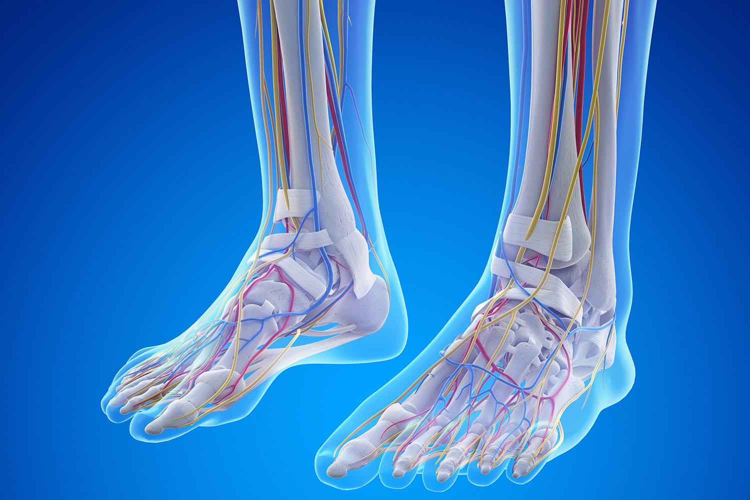
The human feet are a complex structure composed of numerous bones, muscles, tendons, and ligaments that work together to provide support, balance, and mobility. Understanding the different feet parts names and their functions can offer insight into how our feet carry us through life’s journey. Let’s delve into the anatomy of the feet and explore its various components.
The Basic Structure of the Feet
The Feet are divided into three main sections: the forefoot, the midfoot, and the hindfoot. Each section contains specific structures that play crucial roles in foot mechanics.
1. The Forefoot
- Phalanges: These are the bones of the toes. Each toe has three phalanges (proximal, middle, and distal), except for the big toe, which has two (proximal and distal).
- Metatarsals: These long bones connect the phalanges to the midfoot. There are five metatarsals, one for each toe, which provide support and stability.
2. The Midfoot
- Navicular Bone: Located on the inner side of the feet, this bone plays a key role in forming the arch of the feet.
- Cuboid Bone: Situated on the outer side of the feet, the cuboid supports the feet’s lateral side.
- Cuneiform Bones: There are three cuneiform bones (medial, intermediate, and lateral) situated between the navicular bone and the first three metatarsals. They help maintain the arch of the feet.
3. The Hindfoot
- Talus: This bone forms the lower part of the ankle joint and connects the foot to the leg.
- Calcaneus: Commonly known as the heel bone, the calcaneus is the largest bone in the feet and provides the foundation for standing and walking.
Key Ligaments and Tendons
Understanding feet parts names extends beyond bones to include ligaments and tendons, which are essential for movement and stability.
- Plantar Fascia: This thick band of tissue runs from the heel to the toes, supporting the arch and absorbing shock.
- Achilles Tendon: The strongest tendon in the body, it connects the calf muscles to the heel bone, enabling walking, running, and jumping.
- Deltoid Ligament: Located on the inner side of the ankle, this ligament provides stability to the ankle joint.
Muscles of the Feet
The muscles of the Feet can be categorized into intrinsic and extrinsic muscles. Intrinsic muscles are located within the feet, while extrinsic muscles originate from the lower leg and insert into the feet.
- Intrinsic Muscles: These include the lumbricals, interossei, and flexor digitorum brevis, which help with toe movement and maintaining balance.
- Extrinsic Muscles: Major extrinsic muscles include the tibialis anterior, tibialis posterior, and peroneus longus, which control feetand ankle movements.
International Research on Feet Anatomy
Global research has significantly enhanced our understanding of Feet anatomy and its implications for health and mobility. Studies on the biomechanics of the Feet have led to advancements in orthotic design and surgical techniques, improving outcomes for individuals with feet disorders.
For instance, research published in the Journal of Foot and Ankle Research highlights the importance of the medial longitudinal arch in shock absorption and weight distribution. Another study from the International Journal of Orthopaedic and Trauma Nursing emphasizes the role of the plantar fascia in maintaining foot health and the potential impact of plantar fasciitis on mobility.
Understanding the feet parts name and their functions is crucial for appreciating the complexity and importance of our feet. From the bones and ligaments to the muscles and tendons, each component plays a vital role in our daily activities. Continued international research is essential for advancing our knowledge and improving treatments for feet-related conditions, ensuring we can all stand, walk, and run with confidence.


