Overview
Fetal echocardiography, commonly known as fetal echo, is a specialized ultrasound technique used to evaluate the heart of an unborn baby. This non-invasive procedure plays a crucial role in prenatal care, allowing healthcare professionals to detect congenital heart defects (CHDs) and other cardiac abnormalities early in pregnancy. Understanding fetal echo, its significance, and the international research supporting its use is vital for expectant parents and medical practitioners alike.
What is Fetal Echo?
Fetal echo is an advanced form of ultrasound that focuses specifically on the fetal heart. Unlike a standard ultrasound, which provides a general view of the developing fetus, a fetal echo offers detailed images of the heart’s structure and function. This procedure is typically performed between 18 and 24 weeks of gestation, a critical period when the heart is sufficiently developed to be assessed accurately.
Why is Fetal Echo Important?
Congenital heart defects are among the most common types of birth defects, affecting approximately 1 in 100 live births globally. Early detection through fetal echo is crucial for several reasons:
- Early Intervention: Identifying heart defects before birth allows for early intervention and planning. This can include arranging for specialized care during delivery and immediate treatment after birth.
- Improved Outcomes: Research has shown that early diagnosis and intervention can significantly improve the outcomes for babies with CHDs. This includes better surgical success rates and overall survival.
- Parental Preparation: Knowing about a potential heart defect in advance gives parents time to prepare emotionally and logistically, ensuring they are ready to provide the necessary care for their child.
International Research on Fetal Echo
Extensive research has been conducted worldwide to evaluate the effectiveness and benefits of fetal echo. Some key findings include:
- Accuracy and Reliability: Studies have demonstrated that fetal echo is highly accurate in diagnosing various types of congenital heart defects. A study published in the Journal of the American College of Cardiology reported that fetal echo had a diagnostic accuracy of over 90% for detecting major CHDs.
- Impact on Outcomes: A meta-analysis published in the International Journal of Pediatrics highlighted that early detection through fetal echo is associated with improved surgical outcomes and reduced mortality rates for newborns with critical heart defects.
- Technological Advancements: Continuous advancements in ultrasound technology have significantly enhanced the quality of fetal echo imaging. Innovations such as three-dimensional (3D) and four-dimensional (4D) echocardiography provide even more detailed views of the fetal heart, aiding in more precise diagnoses.
- Global Utilization: The use of fetal echo is not limited to high-income countries. Research published in Global Health emphasizes the importance of implementing fetal echo in low- and middle-income countries, where the burden of congenital heart defects is often higher due to limited access to prenatal care.
How is Fetal Echo Performed?
Fetal echo is typically performed by a specialized pediatric cardiologist or a trained sonographer. The procedure involves the following steps:
- Preparation: The expectant mother lies on an examination table, and a special gel is applied to her abdomen to enhance the transmission of sound waves.
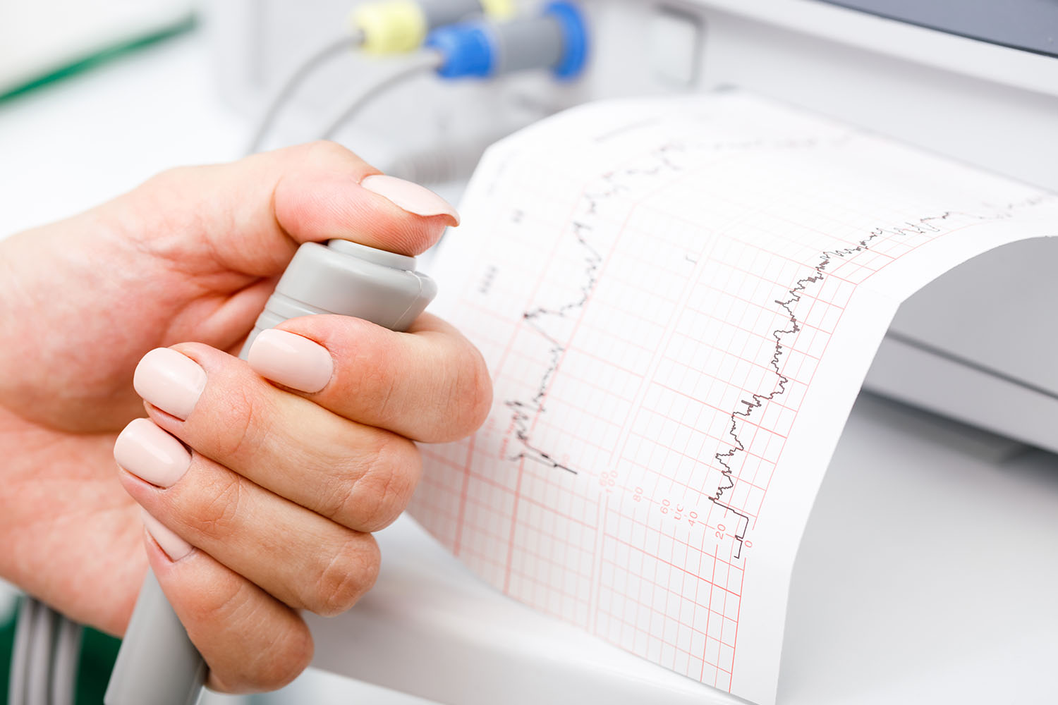
- Ultrasound Imaging: A transducer, a handheld device that emits sound waves, is moved over the abdomen. The sound waves bounce off the fetal heart, creating detailed images on a monitor.
- Analysis: The healthcare provider analyzes the images, assessing the heart’s structure, function, and rhythm. This includes evaluating the chambers, valves, and major blood vessels.
- Discussion: After the procedure, the results are discussed with the parents. If any abnormalities are detected, further tests and consultations with pediatric cardiologists and other specialists may be recommended.
Fetal echo is a vital tool in modern prenatal care, offering early detection and diagnosis of congenital heart defects. The extensive body of international research underscores its accuracy, reliability, and significant impact on improving outcomes for newborns with heart abnormalities. As technology continues to advance, the capabilities of fetal echo will only improve, further enhancing its role in safeguarding the health of unborn children.
For expectant parents, understanding the importance of fetal echo and seeking this specialized ultrasound when recommended can provide peace of mind and ensure the best possible start for their baby’s life.