Overview
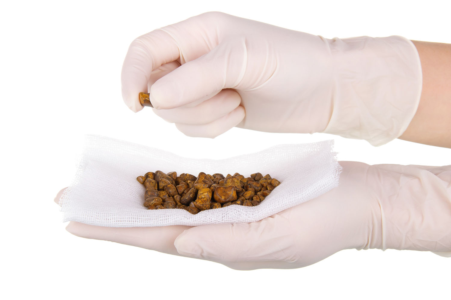
Gallbladder stones, also known as gallstones, are hardened deposits that can form in the gallbladder. These stones can vary in size and number, and their presence can lead to a variety of health issues. In this blog, we will explore the significance of gallbladder stone images, how they are used in diagnosis and treatment, and what international research reveals about this condition.

What are Gallbladder Stones?
Gallbladder stones are typically composed of cholesterol, bilirubin, or a combination of the two. They can range in size from as small as a grain of sand to as large as a golf ball. Gallstones can be asymptomatic or cause severe pain and complications such as inflammation, infection, or blockage of the bile ducts.
Importance of Gallbladder Stone Images
Gallbladder stone images play a crucial role in the medical field for several reasons:
- Diagnosis: Imaging techniques such as ultrasound, CT scans, and MRIs are essential for diagnosing gallstones. These images allow healthcare providers to detect the presence, size, and number of stones within the gallbladder.
- Treatment Planning: Once gallstones are identified, images help in planning the appropriate treatment. For instance, the size and location of the stones can determine whether a patient requires medication, non-invasive procedures, or surgery.
- Monitoring: Gallbladder stone images are also used to monitor the condition over time, especially if the patient is under a non-surgical treatment plan. Regular imaging can help track the effectiveness of the treatment and detect any new stone formation.
Types of Imaging Techniques
- Ultrasound: The most common and preferred method for detecting gallstones. Ultrasound uses sound waves to create images of the gallbladder and can accurately identify the presence of stones.
- CT Scan: Provides detailed cross-sectional images of the body’s internal structures, including the gallbladder. CT scans are often used when complications such as infection or blockage are suspected.
- MRI: Magnetic Resonance Imaging offers high-resolution images and is particularly useful for identifying gallstones in the bile ducts.
International Research on Gallbladder Stones
Research across the globe has provided valuable insights into the causes, prevalence, and treatment of gallstones. Here are some key findings:
- Prevalence: Studies indicate that gallstones are more common in women than in men and that the risk increases with age. Obesity, rapid weight loss, and certain dietary factors also contribute to the formation of gallstones.
- Genetic Factors: Research has shown that genetics can play a significant role in an individual’s susceptibility to developing gallstones. Specific genetic markers have been identified that increase the risk.
- Non-Surgical Treatments: Advances in medicine have led to the development of drugs that can dissolve cholesterol gallstones. However, these treatments are only effective for small stones and may take months or years to work.
- Surgical Interventions: Laparoscopic cholecystectomy, a minimally invasive surgery to remove the gallbladder, is the most common and effective treatment for symptomatic gallstones. This procedure has a high success rate and a relatively quick recovery time.
Gallbladder stone images are invaluable tools in the diagnosis, treatment, and management of gallstones. With advances in imaging technology and ongoing international research, healthcare providers can offer more accurate diagnoses and effective treatments for those affected by this condition. Understanding and utilizing gallbladder stone images can lead to better patient outcomes and a deeper comprehension of this common yet potentially serious health issue.


