Overview
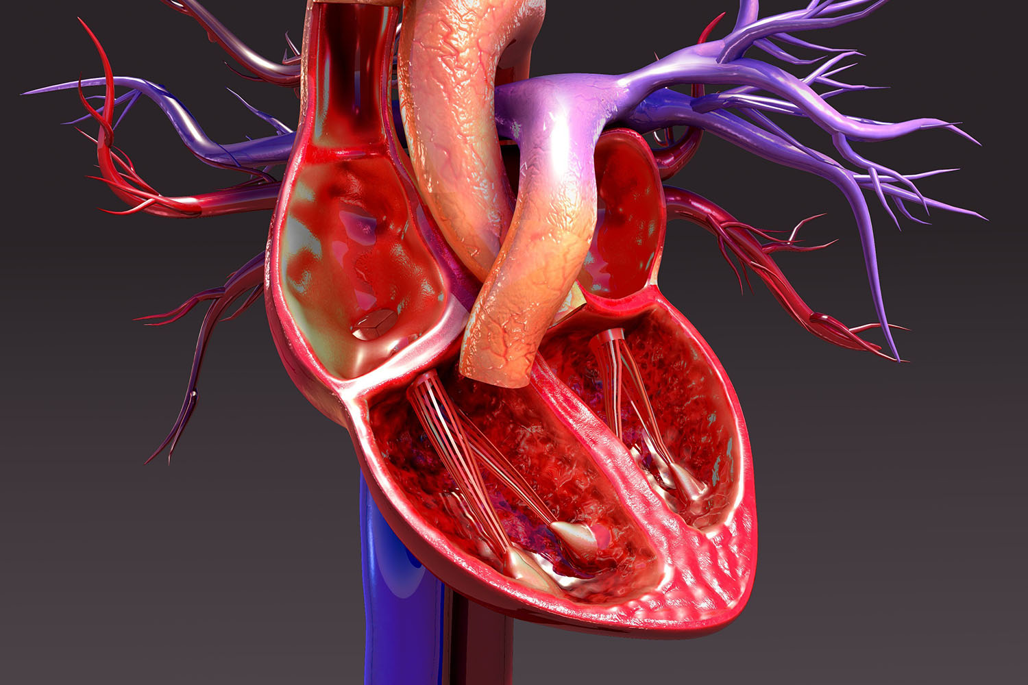
Understanding the heart’s intricate workings is crucial for diagnosing and treating various cardiovascular conditions. The field of cardiac imaging has evolved significantly, providing us with clear, detailed heart images in the human body, allowing for precise analysis and intervention.
The Importance of Cardiac Imaging
Cardiac imaging plays a pivotal role in modern medicine. It helps in the early detection of heart diseases, planning and monitoring treatments, and improving patient outcomes. With heart diseases being a leading cause of mortality globally, the advancements in imaging technology are vital for better healthcare.
Types of Cardiac Imaging Techniques
Several imaging techniques are used to capture detailed heart images in the human body, each with unique benefits and applications. Here are some of the most commonly used methods:
1. Echocardiography
Echocardiography uses ultrasound waves to create detailed images of the heart. It is widely used due to its non-invasive nature and ability to provide real-time images of the heart’s structure and function. This technique is essential for evaluating heart valves, chambers, and overall cardiac performance.
2. Magnetic Resonance Imaging (MRI)
Cardiac MRI is a sophisticated imaging technique that offers high-resolution images of the heart. It provides detailed information about the heart’s anatomy, function, and tissue characteristics. MRI is particularly useful for detecting myocardial infarctions, cardiomyopathies, and congenital heart defects.
3. Computed Tomography (CT)
CT scans produce detailed cross-sectional images of the heart and blood vessels. Coronary CT angiography (CTA) is a specific type of CT scan that visualizes the coronary arteries, helping to detect blockages or narrowing. CT is also used to assess cardiac masses and aortic diseases.
4. Nuclear Cardiology
Nuclear cardiology involves the use of radioactive tracers to create images of the heart. Techniques such as Single Photon Emission Computed Tomography (SPECT) and Positron Emission Tomography (PET) provide functional information about blood flow and myocardial viability, which are crucial for diagnosing coronary artery disease and assessing the extent of myocardial damage.
5. Electrocardiogram (ECG) Imaging
While not an imaging technique in the traditional sense, ECG imaging records the electrical activity of the heart and is often used alongside other imaging modalities to provide a comprehensive view of cardiac health. It helps in diagnosing arrhythmias, myocardial infarctions, and other electrical abnormalities.
International Research and Advances
The global research community has made significant strides in advancing cardiac imaging technologies. Here are some noteworthy developments:
Artificial Intelligence in Cardiac Imaging
Artificial Intelligence (AI) is revolutionizing cardiac imaging by enhancing image analysis, improving diagnostic accuracy, and predicting patient outcomes. AI algorithms can quickly analyze large sets of data, identify subtle patterns, and assist radiologists in making precise diagnoses. For instance, a study published in The Lancet demonstrated that AI could accurately detect heart disease from cardiac MRI images, potentially reducing the workload on clinicians and improving diagnostic speed.
3D Printing and Heart Models
3D printing technology is being used to create precise anatomical models of the heart based on imaging data. These models are invaluable for surgical planning, education, and patient communication. Surgeons can practice complex procedures on 3D-printed hearts, leading to better surgical outcomes and reduced operative times.
Molecular Imaging
Molecular imaging techniques are being developed to provide insights into the biological processes at the molecular level within the heart. These techniques allow for the early detection of disease, monitoring of treatment responses, and the development of personalized therapies. Research published in Nature Reviews Cardiology highlighted the potential of molecular imaging in identifying inflammatory processes in atherosclerosis, which could lead to more targeted and effective treatments.
Global Collaborations
International collaborations among research institutions, medical centers, and industry leaders are driving innovation in cardiac imaging. Collaborative efforts like the Global Burden of Disease Study provide valuable data on cardiovascular health trends worldwide, informing public health policies and research priorities.
The advancements in cardiac imaging are transforming the way we understand and treat heart diseases. With ongoing research and technological innovations, the future of heart images in the human body looks promising, offering hope for better diagnosis, treatment, and prevention of cardiovascular conditions. As we continue to explore the depths of the human heart, these images will remain a cornerstone of cardiac health, guiding us towards a healthier future.


