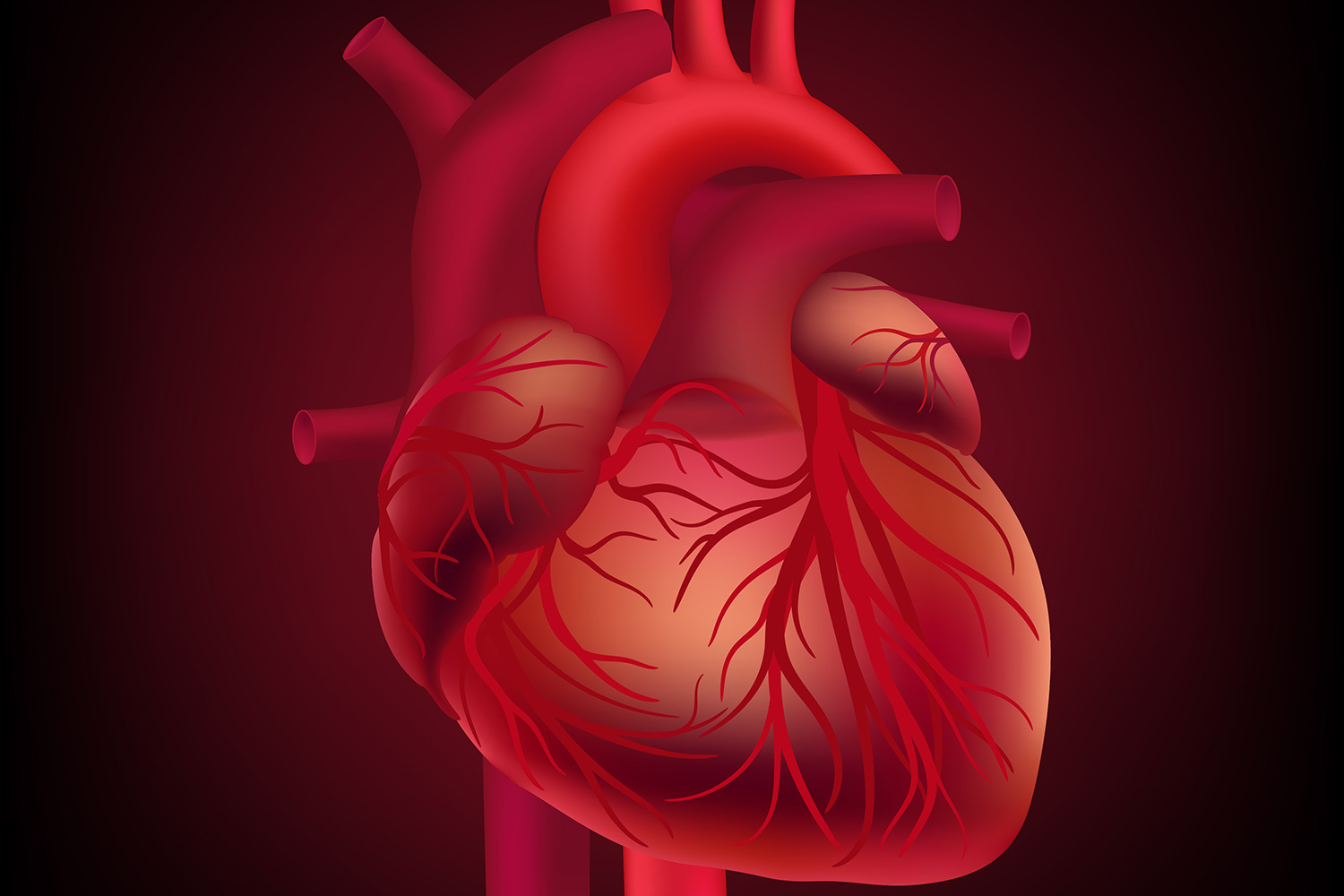
The heart, a crucial organ in the circulatory system, is a marvel of biological engineering. Its intricate design ensures the effective circulation of blood throughout the body, delivering oxygen and nutrients while removing waste products. Understanding the internal structure of the heart is fundamental to comprehending its function and diagnosing various heart conditions. This blog delves into the detailed anatomy of the heart, supported by international research and visual aids.
Anatomy of the Heart
The heart is a muscular organ roughly the size of a fist, located slightly left of the center of the chest. It consists of four chambers and several key structures that work in unison to pump blood efficiently. The internal structure of the heart diagram provides a comprehensive view of these components:
1. Chambers of the Heart
- Right Atrium: Receives deoxygenated blood from the body via the superior and inferior vena cava. The blood is then transferred to the right ventricle.
- Right Ventricle: Pumps deoxygenated blood through the pulmonary artery to the lungs for oxygenation.
- Left Atrium: Receives oxygenated blood from the lungs via the pulmonary veins. This blood is then transferred to the left ventricle.
- Left Ventricle: Pumps oxygen-rich blood through the aorta to the rest of the body.
2. Valves of the Heart
The heart contains four main valves that maintain unidirectional blood flow:
- Tricuspid Valve: Located between the right atrium and right ventricle.
- Pulmonary Valve: Situated between the right ventricle and the pulmonary artery.
- Mitral Valve: Positioned between the left atrium and left ventricle.
- Aortic Valve: Found between the left ventricle and the aorta.
3. Septum
The septum is a muscular wall that divides the heart into its right and left sides, preventing the mixing of oxygenated and deoxygenated blood.
4. Heart Wall Layers
The heart wall comprises three layers:
- Epicardium: The outer layer, which also serves as the heart’s protective covering.
- Myocardium: The middle layer, consisting of cardiac muscle tissue responsible for heart contractions.
- Endocardium: The inner layer, which lines the chambers and valves.
Internal Structure of the Heart Diagram
An internal structure of the heart diagram offers a visual representation of the heart’s anatomy, making it easier to understand the spatial relationships between its components. The diagram typically highlights:
- The four chambers (right atrium, right ventricle, left atrium, left ventricle).
- The four valves (tricuspid, pulmonary, mitral, aortic).
- The septum and the major blood vessels (aorta, pulmonary arteries, vena cavae, pulmonary veins).
International Research and Clinical Relevance
International research has significantly advanced our understanding of the heart’s internal structure. Studies have utilized advanced imaging techniques, such as MRI and echocardiography, to create detailed and accurate heart diagrams. These technologies have improved the diagnosis and treatment of cardiovascular diseases by providing insights into the heart’s structure and function.
For instance, research has shown that abnormalities in the internal structure of the heart, such as valve defects or septal defects, can lead to serious health issues. Early detection through imaging can help in managing these conditions effectively.
A detailed internal structure of the heart diagram is a valuable tool for both medical professionals and students. It offers a clear view of how the heart functions and helps in diagnosing and treating heart-related conditions. Understanding the heart’s anatomy is crucial for appreciating its role in maintaining overall health and well-being. As research continues to evolve, our knowledge of the heart’s structure and its implications for health will only become more precise and informative.


