Overview
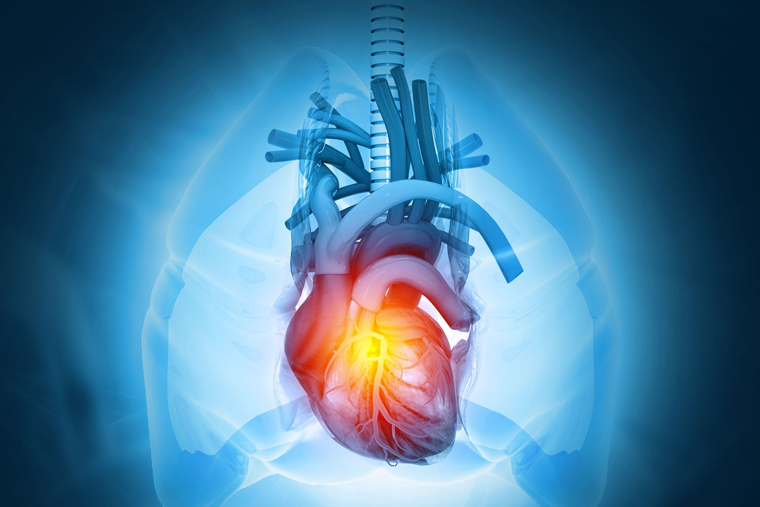
The human heart, a vital organ responsible for pumping blood throughout the body, has a complex and intricate internal structure that ensures efficient circulation. Understanding the internal structure of the human heart is crucial for comprehending how this essential organ functions.
The Chambers of the Heart
The heart is divided into four chambers: two atria and two ventricles. The atria are the upper chambers, and the ventricles are the lower chambers. These chambers work together to pump blood throughout the body.
Atria
- Right Atrium: Receives deoxygenated blood from the body through the superior and inferior vena cava.
- Left Atrium: Receives oxygenated blood from the lungs through the pulmonary veins.
Ventricles
- Right Ventricle: Pumps deoxygenated blood to the lungs via the pulmonary artery.
- Left Ventricle: Pumps oxygenated blood to the rest of the body through the aorta.
Heart Valves
The heart contains four main valves that ensure the unidirectional flow of blood and prevent backflow. These valves are:
Atrioventricular (AV) Valves
- Tricuspid Valve: Located between the right atrium and right ventricle.
- Mitral Valve (Bicuspid Valve): Located between the left atrium and left ventricle.
Semilunar Valves
- Pulmonary Valve: Located between the right ventricle and the pulmonary artery.
- Aortic Valve: Located between the left ventricle and the aorta.
The Cardiac Conduction System
The internal structure of the human heart includes a specialized conduction system that controls the heart’s rhythm and ensures synchronized contractions. The main components of this system are:
Sinoatrial (SA) Node
- Often referred to as the heart’s natural pacemaker, the SA node initiates electrical impulses that cause the atria to contract.
Atrioventricular (AV) Node
- Receives the impulse from the SA node and delays it slightly to allow the ventricles to fill with blood before contracting.
Bundle of His
- Transmits impulses from the AV node to the ventricles.
Purkinje Fibers
- Distribute the electrical impulse throughout the ventricles, causing them to contract.
The Coronary Circulation
The coronary circulation is a crucial aspect of the internal structure of the human heart. It provides the heart muscle (myocardium) with the necessary oxygen and nutrients to function effectively. The coronary arteries, which branch off from the aorta, supply oxygen-rich blood to the heart muscle, while the coronary veins remove deoxygenated blood.
The Myocardium
The myocardium is the thick, muscular layer of the heart wall responsible for the contractile function of the heart. It is composed of specialized cardiac muscle cells that can conduct electrical impulses and contract in a coordinated manner.
The Endocardium and Epicardium
The heart wall has three layers:
- Endocardium: The innermost layer, lining the chambers and valves.
- Myocardium: The middle, muscular layer.
- Epicardium: The outermost layer, which forms part of the pericardium, a double-walled sac containing the heart.
The internal structure of the human heart is a marvel of biological engineering, consisting of chambers, valves, a conduction system, and a dedicated coronary circulation. Each component works in harmony to ensure that blood is efficiently pumped throughout the body, maintaining the essential functions of life. Understanding this intricate structure helps us appreciate the heart’s role in sustaining our health and well-being.


