Overview
When discussing spinal anatomy and various medical procedures involving the spine, the term “pedicle” frequently arises. The pedicle is a small but significant structure, playing an essential role in both the stability and function of the vertebral column. This blog explores the anatomy, function, and clinical significance of the pedicle, drawing on international research to highlight its importance in spinal health and surgery.
Anatomy of the Pedicle
The pedicle is a stub of bone that connects the lamina to the vertebral body in each vertebra of the spinal column. It forms part of the vertebral arch and is located posteriorly to the vertebral body. Each vertebra has two pedicles, one on each side, creating a bridge-like structure that provides robust support and protection for the spinal cord and nerves.
Function of the Pedicle
The primary functions of the pedicle include:
- Structural Support: The pedicles bear significant loads and transmit forces from the vertebral body to the posterior elements of the spine, such as the lamina and spinous processes.
- Protection: They form the walls of the vertebral foramen, through which the spinal cord passes, providing a protective bony encasement.
- Anchorage for Muscles and Ligaments: Numerous muscles and ligaments attach to the pedicles, contributing to spinal stability and flexibility.
Clinical Significance of the Pedicle
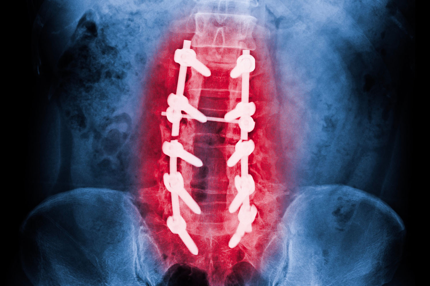
Pedicle Screw Fixation
One of the most critical applications of the pedicle in clinical practice is in pedicle screw fixation. This surgical technique is widely used in spinal fusion procedures to stabilize the spine. Pedicle screws are inserted into the vertebrae’s pedicles and connected by rods to immobilize and support the spine as it heals. This method is particularly effective for treating conditions such as scoliosis, spondylolisthesis, and spinal fractures.
Research has shown that pedicle screw fixation provides superior stability compared to other spinal instrumentation methods. A study published in the Journal of Spinal Disorders & Techniques demonstrated that this technique significantly enhances fusion rates and improves clinical outcomes in patients with various spinal pathologies .
Pedicle Morphometry
Understanding the morphometry of the pedicle is crucial for the safe and effective placement of pedicle screws. Variations in pedicle size and shape can occur between individuals and across different populations. A study in the European Spine Journal highlighted the importance of considering ethnic differences in pedicle dimensions when planning spinal surgeries . This research underscores the need for individualized preoperative planning using imaging techniques like CT scans to avoid complications such as pedicle breaches or nerve damage.
Pedicle Pathologies
Pedicle Fractures
Pedicle fractures, though less common than other vertebral fractures, can occur due to trauma or as a result of underlying conditions like osteoporosis. These fractures can compromise spinal stability and may require surgical intervention to prevent further damage and alleviate pain.
Pedicle Lysis
Pedicle lysis refers to the dissolution or weakening of the pedicle, often seen in conditions like spondylolysis. This can lead to vertebral slippage (spondylolisthesis), causing pain and neurological deficits. Early detection and appropriate management are crucial to prevent progression and improve patient outcomes.
Advances in Pedicle Research
Ongoing research continues to enhance our understanding of the pedicle and its applications in spinal surgery. Recent advancements include the development of 3D printing technology for creating patient-specific pedicle screw guides, improving the accuracy and safety of screw placement. Additionally, biomechanical studies are exploring new materials and designs for pedicle screws to further enhance spinal stability and reduce the risk of hardware failure.
Conclusion
The pedicle, though small in size, plays a vital role in the structural integrity and function of the spine. Its importance in spinal surgery, particularly in pedicle screw fixation, highlights the need for a thorough understanding of its anatomy and variations. Ongoing research and technological advancements continue to improve our ability to treat spinal conditions effectively, ensuring better outcomes for patients worldwide.
By delving into the complexities of the pedicle, we gain a deeper appreciation for this critical component of spinal anatomy and its significance in maintaining spinal health and facilitating successful surgical interventions.