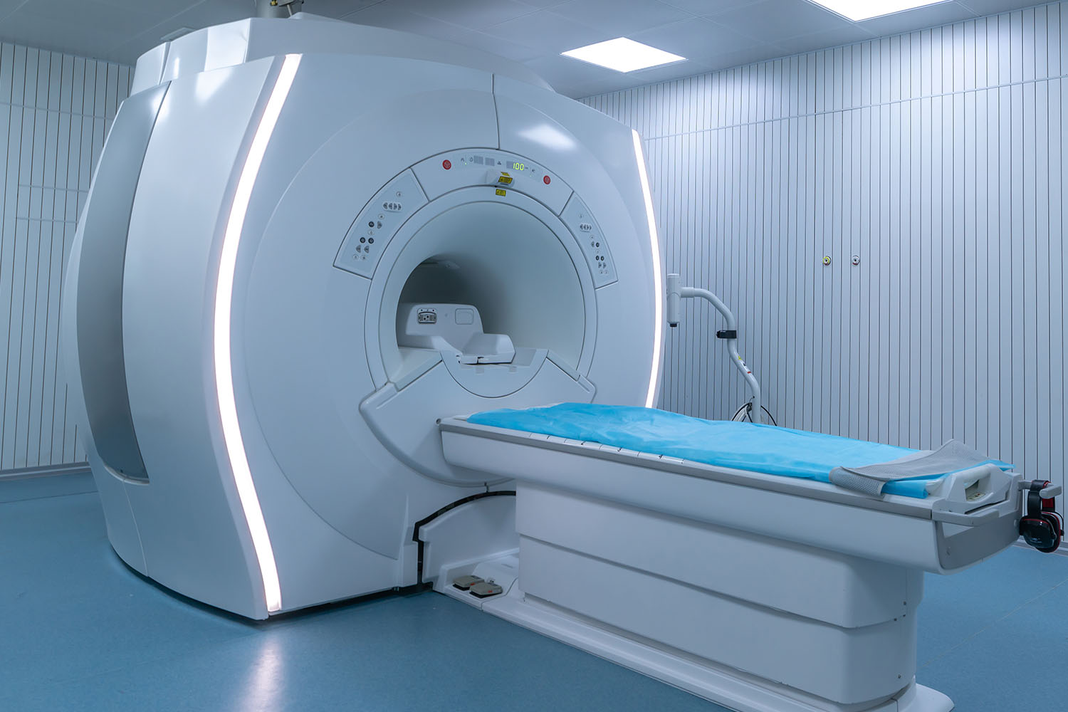
Overview
Positron emission tomography, commonly abbreviated as PET, is a powerful and sophisticated imaging technique used in the fields of medical diagnostics and research. The name itself reveals much about the process: positrons are the positively charged counterparts of electrons, and tomography refers to imaging by sections. This blog delves into the full form of PET, its research applications, and some intriguing facts about this technology.

What is Positron Emission Tomography?
Positron emission tomography is an imaging modality that helps visualize the metabolic processes in the body. Unlike conventional imaging techniques such as X-rays or MRI, which primarily provide structural information, PET scans offer insights into the functional aspects of organs and tissues. This makes PET particularly valuable in detecting and monitoring diseases that alter cellular activity, such as cancer, heart disease, and neurological disorders.
How Does Positron Emission Tomography Work?
The process begins with the injection of a radiotracer, a substance labeled with a positron-emitting radionuclide. One of the most commonly used radiotracers is fluorodeoxyglucose (FDG), which is similar to glucose and is preferentially taken up by metabolically active cells. Once inside the body, the radiotracer emits positrons that collide with electrons, resulting in the emission of gamma rays. These gamma rays are detected by the PET scanner, which constructs detailed images reflecting the concentration and distribution of the radiotracer.
Research Applications of Positron Emission Tomography
Cancer Diagnosis and Treatment: PET scans are extensively used in oncology to detect tumors, evaluate the extent of cancer spread (staging), and monitor the effectiveness of treatment. The ability of PET to distinguish between benign and malignant lesions is crucial for early and accurate diagnosis.
Neurology: Positron emission tomography plays a significant role in understanding brain function and diagnosing neurological conditions such as Alzheimer’s disease, epilepsy, and Parkinson’s disease. By highlighting areas of altered metabolic activity, PET scans can reveal the presence and progression of these disorders.
Cardiology: In cardiology, PET scans are used to assess myocardial perfusion and viability. This helps in identifying areas of the heart muscle that are still viable after a heart attack and determining the best treatment strategy.
Pharmaceutical Research: PET is a valuable tool in drug development and pharmacokinetics, providing insights into how new drugs are absorbed, distributed, and metabolized in the body. This information is critical for optimizing drug formulations and dosages.
Fascinating Facts About Positron Emission Tomography
Historical Development: The concept of positron emission tomography dates back to the 1950s and 1960s, with the first PET scanner developed in the 1970s. The technique has since evolved, becoming an indispensable tool in modern medicine.
Combining Modalities: PET scans are often combined with computed tomography (CT) scans, resulting in PET/CT, which provides both metabolic and anatomical information in a single session. This combination enhances diagnostic accuracy and patient management.
Radiotracer Innovations: While FDG is the most common radiotracer, researchers are continually developing new tracers to target specific cellular processes and receptors. These innovations expand the potential applications of PET in various medical fields.
Non-Invasive and Painless: One of the significant advantages of positron emission tomography is its non-invasive nature. Patients undergo a simple injection followed by a scan, making it a relatively straightforward and painless procedure.
Advances in Technology: Technological advancements are continually improving PET scanner resolution, sensitivity, and speed. These improvements enhance image quality and reduce scan times, making the process more efficient and comfortable for patients.
Positron emission tomography stands at the forefront of medical imaging, offering unparalleled insights into the body’s metabolic and physiological processes. Its contributions to cancer diagnosis, neurological research, cardiology, and pharmaceutical development underscore its importance in modern healthcare. As technology advances, the capabilities and applications of positron emission tomography will continue to expand, paving the way for even more significant breakthroughs in medicine.


