Overview
The radius bone, one of the two primary bones in the forearm, plays a crucial role in the functionality and movement of the arm. Located on the lateral side of the forearm, extending from the elbow to the wrist, the radius is pivotal in enabling complex motions such as rotation, lifting, and bending. This blog delves into the intricate details of radius bone anatomy, its functions, and its clinical significance, drawing on international research to provide a comprehensive overview.
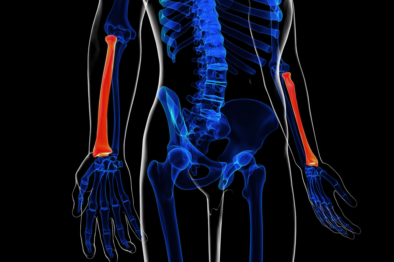
Basic Structure of the Radius Bone
The radius bone is divided into three main parts: the proximal end, the shaft, and the distal end.
- Proximal End: This part of the radius is closest to the elbow. It features the radial head, a disc-shaped structure that articulates with the capitulum of the humerus and the radial notch of the ulna, allowing for the rotation of the forearm. The radial neck connects the head to the radial tuberosity, a bony prominence that serves as an attachment point for the biceps brachii muscle.
- Shaft: The shaft of the radius is the long, central portion of the bone. It is slightly curved and has a triangular cross-section. The interosseous border, located along the medial side of the shaft, is where the interosseous membrane attaches, linking the radius and ulna and providing stability to the forearm.
- Distal End: This end of the radius is closest to the wrist. It is broader and articulates with the carpal bones of the wrist, specifically the scaphoid and lunate bones, forming part of the wrist joint. The distal end also features the styloid process, a bony projection that provides attachment for ligaments of the wrist.
Functions of the Radius Bone
The radius bone plays several vital roles in the arm’s functionality:
- Articulation and Movement: The radius, in conjunction with the ulna, allows for the rotation of the forearm (pronation and supination). This movement is essential for various daily activities, such as turning a doorknob or using a screwdriver.
- Support and Stability: The radius provides structural support to the forearm, helping to maintain its shape and integrity. The interosseous membrane connecting the radius and ulna adds stability and facilitates the transfer of forces between the two bones.
- Muscle Attachment: Numerous muscles and tendons attach to the radius, contributing to the movement and strength of the forearm and hand.
Clinical Significance of Radius Bone Anatomy
Understanding the radius bone anatomy is crucial in diagnosing and treating various medical conditions and injuries. Some common issues include:
- Fractures: Radius fractures, particularly distal radius fractures, are common injuries, especially among older adults. Proper understanding of radius bone anatomy aids in effective treatment, which may involve casting, splinting, or surgical intervention.
- Dislocations and Ligament Injuries: The radius can be involved in dislocations and ligament injuries, particularly at the wrist joint. Knowledge of the bone’s anatomy helps in accurate diagnosis and appropriate management.
- Developmental and Congenital Disorders: Conditions such as radial dysplasia, where the radius bone is underdeveloped or absent, require a thorough understanding of normal radius anatomy for effective surgical planning and intervention.
Insights from International Research
Recent international studies have provided deeper insights into radius bone anatomy and its implications in medical practice. Research from the Journal of Hand Surgery and the International Journal of Orthopaedic Surgery highlights advancements in surgical techniques and rehabilitation protocols for radius fractures. Studies emphasize the importance of precise anatomical knowledge in improving patient outcomes and reducing complications.
For instance, a study published in The Lancet demonstrated the efficacy of new plating techniques for distal radius fractures, showing significantly better functional recovery and fewer complications compared to traditional methods. Another research article from the Journal of Anatomy explored the variations in radius bone morphology among different populations, underlining the importance of considering anatomical diversity in clinical practice.
The radius bone anatomy is integral to the functionality and movement of the forearm and hand. A thorough understanding of its structure and functions is essential for diagnosing and treating various conditions affecting the forearm. Ongoing international research continues to enhance our knowledge, leading to better clinical practices and improved patient care. Whether you are a medical professional, a student, or simply someone interested in human anatomy, the radius bone’s intricate design and critical role in daily activities make it a fascinating subject to explore.