Overview
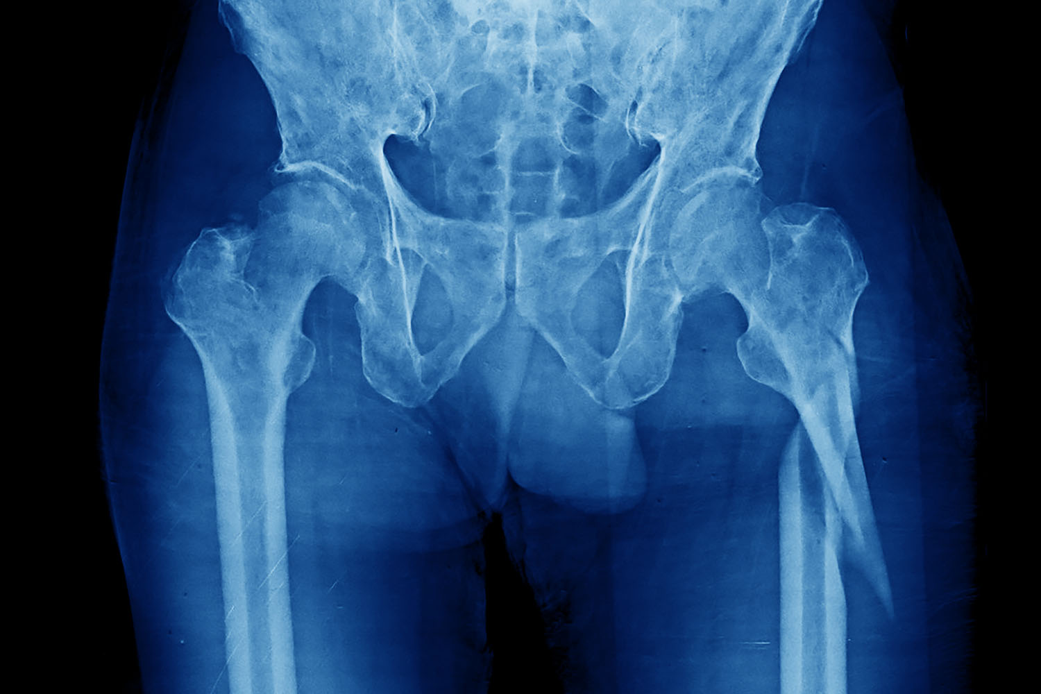
A subtrochanteric fracture refers to a break in the femur (thigh bone) just below the lesser and greater trochanters, which are bony prominences near the hip joint. This type of fracture is significant due to its location and the high stress placed on this part of the femur. Understanding the causes, treatment options, and ongoing research is crucial for medical professionals and patients alike.
Causes and Risk Factors
Subtrochanteric fractures are often the result of high-energy trauma, such as car accidents or falls from significant heights. In elderly patients, these fractures can also occur due to low-energy trauma, especially in those with weakened bones from osteoporosis. Other risk factors include:
- Age: Elderly individuals are more susceptible due to decreased bone density.
- Gender: Women are at higher risk, particularly post-menopausal women due to osteoporosis.
- Medications: Long-term use of corticosteroids can weaken bones.
- Genetic Factors: Certain genetic conditions can predispose individuals to weaker bones.
Symptoms and Diagnosis
Common symptoms of a subtrochanteric fracture include:
- Severe pain in the hip or thigh.
- Inability to bear weight on the affected leg.
- Swelling and bruising in the hip or thigh area.
- Deformity of the leg.
Diagnosis typically involves a physical examination followed by imaging studies. X-rays are the primary diagnostic tool, but CT scans or MRIs may be used for more detailed visualization of the fracture and surrounding tissues.
Treatment Options
Treatment of subtrochanteric fractures generally involves surgical intervention, as conservative treatment is rarely effective due to the high stresses on this part of the femur. The main surgical options include:
- Intramedullary Nailing: This is the most common surgical treatment. A metal rod is inserted into the marrow canal of the femur to stabilize the bone.
- Plate Fixation: In cases where intramedullary nailing is not suitable, a metal plate is attached to the outside of the bone using screws.
- External Fixation: In severe cases with extensive soft tissue damage, external fixation may be used temporarily until the patient is stable enough for a more permanent solution.
Rehabilitation and Recovery
Post-surgery, rehabilitation is crucial for recovery. Physical therapy focuses on restoring mobility, strength, and function. The timeline for recovery can vary, but most patients can expect to begin weight-bearing activities within a few weeks post-surgery, with full recovery taking several months.
International Research and Advances
Recent international research has focused on improving the outcomes of subtrochanteric fracture treatments. Some notable areas of study include:
- Biomechanical Studies: Research on the optimal design and placement of intramedullary nails and plates to enhance stability and promote faster healing.
- Minimally Invasive Techniques: Advances in surgical techniques that reduce soft tissue damage, thereby decreasing recovery time and improving outcomes.
- Bone Healing Agents: Studies on pharmacological agents that can promote bone healing, such as bone morphogenetic proteins (BMPs) and parathyroid hormone (PTH) analogs.
- Osteoporosis Management: Research on better management of osteoporosis to prevent fractures, including the use of new medications and lifestyle interventions.
Subtrochanteric fractures are serious injuries that require prompt and effective treatment to ensure the best possible outcomes. With advances in surgical techniques and ongoing research into bone healing and osteoporosis management, the prognosis for patients with these fractures continues to improve. Understanding the causes, symptoms, and treatment options, as well as staying informed about the latest research, is essential for both healthcare providers and patients dealing with subtrochanteric fractures.


