Overview
The human body comprises various types of joints, each serving unique functions. Among these is the syndesmosis joint, a fibrous joint where two bones are connected by a ligament. This blog explores the syndesmosis joint, delving into its anatomy, function, common injuries, and treatments, supported by international research.
Anatomy of the Syndesmosis Joint
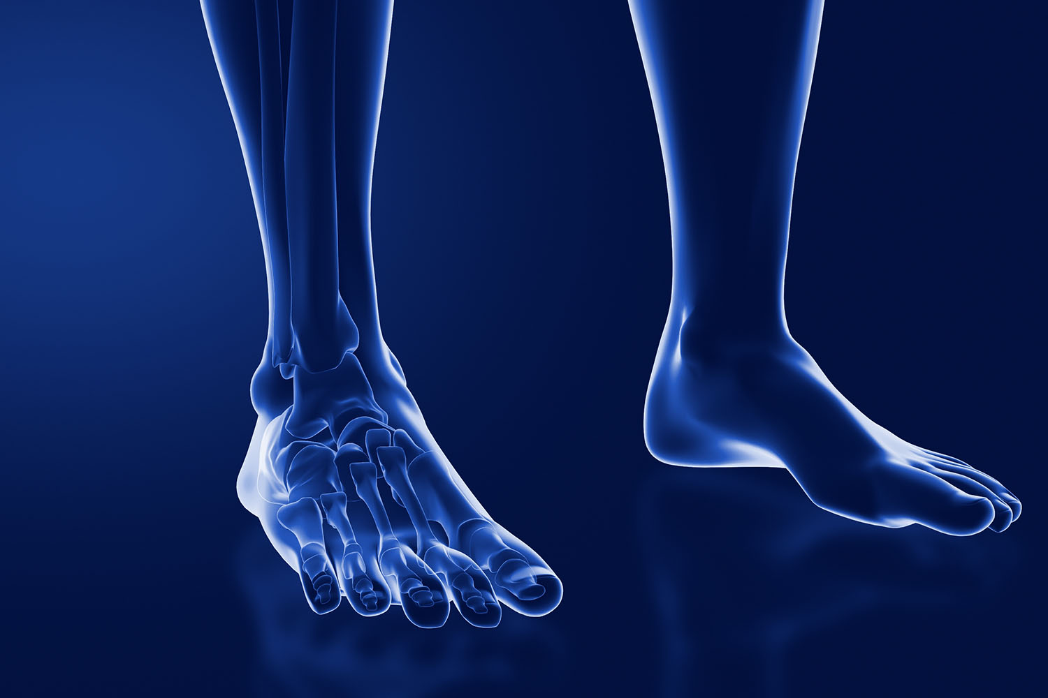
The syndesmosis joint is found between the tibia and fibula in the lower leg, as well as between the radius and ulna in the forearm. Unlike synovial joints, which allow for significant movement, syndesmosis joints are relatively immobile. They are classified as amphiarthroses, meaning they permit slight movement. The primary structure connecting the bones in a syndesmosis joint is the interosseous membrane, a dense fibrous tissue providing stability and distributing forces across the joint.
Function of the Syndesmosis Joint
The primary function of the syndesmosis joint is to maintain the proper alignment of adjacent bones while allowing for limited movement. In the lower leg, the tibiofibular syndesmosis plays a crucial role in stabilizing the ankle joint and enabling efficient transmission of weight from the leg to the foot. The radioulnar syndesmosis in the forearm allows for the rotation of the forearm, a movement essential for various daily activities.
Common Injuries
Injuries to the syndesmosis joint, often referred to as syndesmotic injuries, are prevalent in athletes and individuals involved in high-impact activities. The most common injury is a high ankle sprain, which occurs when the ligaments connecting the tibia and fibula are torn or stretched. Unlike typical ankle sprains, which involve the ligaments on the outside of the ankle, high ankle sprains affect the ligaments above the ankle joint, making them more severe and requiring a longer recovery period.
Diagnosis and Treatment
Diagnosing syndesmosis joint injuries involves a combination of physical examinations and imaging techniques such as X-rays, MRI, or CT scans. The physical examination often includes specific tests like the squeeze test or external rotation test to assess the integrity of the syndesmotic ligaments.
Treatment for syndesmosis joint injuries varies depending on the severity of the injury. Mild injuries may be treated with rest, ice, compression, and elevation (RICE), along with physical therapy to restore strength and mobility. More severe injuries might require surgical intervention to repair or reconstruct the damaged ligaments. International research emphasizes the importance of early and accurate diagnosis to prevent long-term complications such as chronic instability or arthritis.
International Research and Perspectives
Research from around the world has significantly contributed to our understanding of the syndesmosis joint. A study published in the Journal of Orthopaedic Research highlighted the biomechanical properties of the syndesmosis joint, emphasizing the role of the interosseous membrane in maintaining joint stability. Another study from the British Journal of Sports Medicine focused on the rehabilitation protocols for syndesmotic injuries, providing evidence-based guidelines for effective recovery.
The syndesmosis joint, though less commonly discussed than other joints, plays a vital role in the stability and function of the lower leg and forearm. Understanding its anatomy, function, and the impact of injuries is crucial for proper diagnosis and treatment. Continued international research is essential to advance our knowledge and improve outcomes for individuals with syndesmotic injuries.
In summary, whether you are an athlete, a healthcare professional, or someone interested in human anatomy, appreciating the importance of the syndesmosis joint can enhance your understanding of the complex interplay between the body’s structures.


