Overview
In the realm of cardiology, the echocardiogram stands as a powerful tool, offering insights into the inner workings of the heart with remarkable clarity. This non-invasive procedure, also known as a cardiac echo or simply an echo, utilizes sound waves to create detailed images of the heart’s structure and function. But what exactly does an echocardiogram show, and why is it such a vital diagnostic tool in the world of cardiovascular medicine? Let’s embark on a journey through the chambers of the heart to uncover the answers.
Visualizing the Heart’s Anatomy: A Detailed Portrait
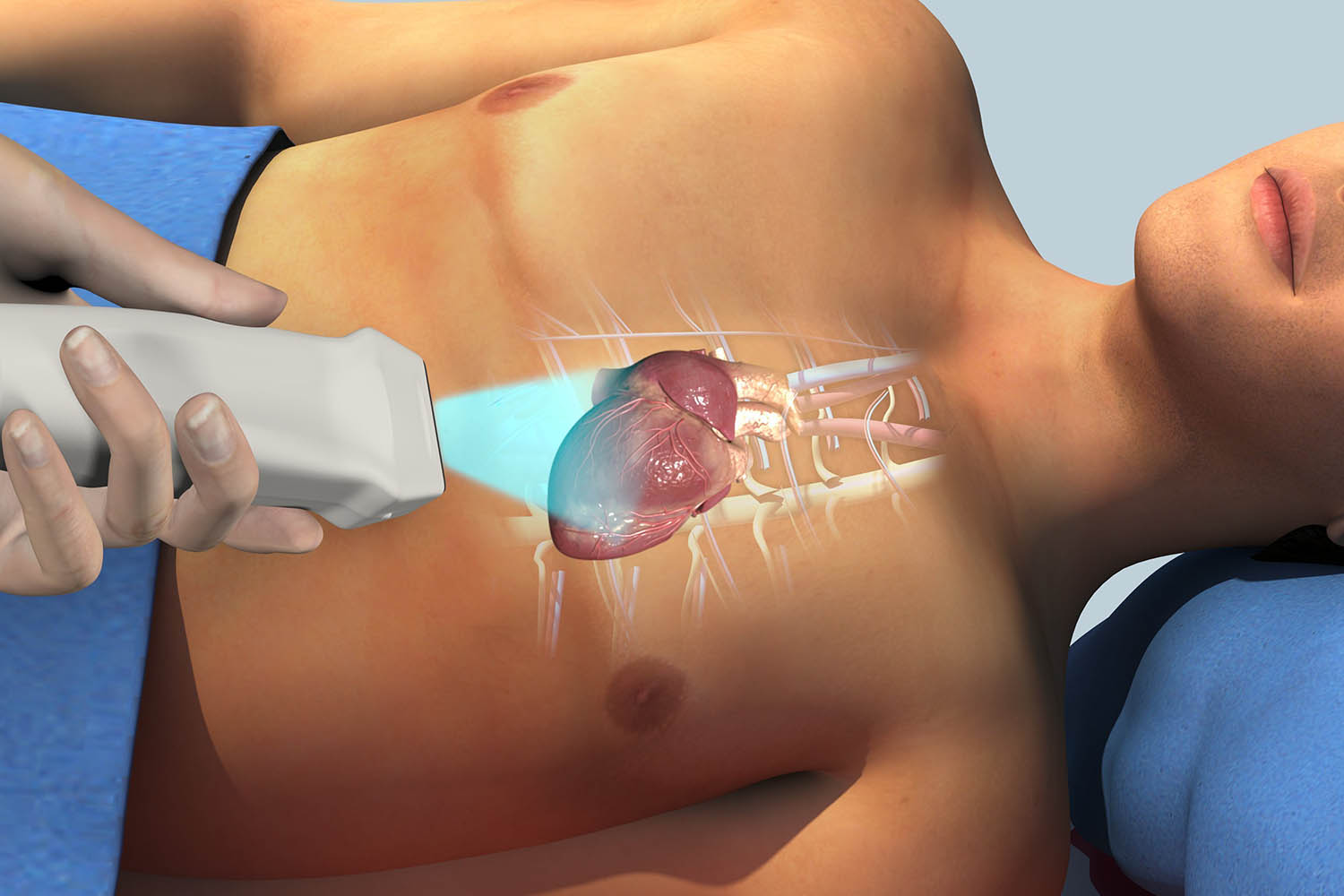
At the core of an echocardiogram lies its ability to provide a comprehensive view of the heart’s anatomy. Using high-frequency sound waves, or ultrasound, the echo captures real-time images of the heart’s chambers, valves, and surrounding structures. These images allow cardiologists to assess the size, shape, and movement of the heart, providing crucial insights into its overall health and function.
Assessing Cardiac Function: The Beat of the Heart
Beyond mere anatomy, an echocardiogram delves into the dynamic aspects of cardiac function. By measuring parameters such as ejection fraction (the percentage of blood pumped out of the heart with each contraction), cardiac output, and wall motion abnormalities, the echo offers valuable information about the heart’s pumping ability and efficiency. This data is essential for diagnosing conditions such as heart failure, cardiomyopathy, and myocardial infarction (heart attack).
Detecting Valve Abnormalities: Gatekeepers of Circulation
The heart’s valves play a crucial role in maintaining the unidirectional flow of blood through its chambers. An echocardiogram can detect abnormalities in valve structure and function, such as stenosis (narrowing), regurgitation (leakage), or prolapse (bulging). These findings aid in the diagnosis and management of conditions such as mitral valve prolapse, aortic stenosis, and mitral regurgitation.
Identifying Structural Anomalies: Unveiling Hidden Defects
In addition to assessing the heart’s main components, an echocardiogram can reveal structural anomalies and congenital heart defects. From atrial septal defects (ASDs) and ventricular septal defects (VSDs) to abnormalities in chamber size and orientation, the echo serves as a critical tool for diagnosing these often-hidden conditions, particularly in pediatric patients.
Monitoring Disease Progression: A Window Into Treatment
Beyond its diagnostic utility, an echocardiogram plays a pivotal role in monitoring disease progression and treatment response. Serial echocardiograms track changes in cardiac function over time, guiding therapeutic interventions and helping clinicians tailor treatment strategies to individual patients’ needs.